Figure 8.

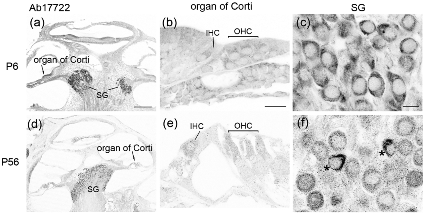
FMRP_Ab17722 immunostaining in the gerbil cochlea during development. (a–c) were taken from a P6 gerbil. (d–f) were taken from a P56 gerbil. (a) and (d) are low-magnification images of the cochlea. (b) and (e) are high-magnification images of the organ of Corti. (c) and (f) were taken from the spiral ganglion (SG). Asterisks in (f) indicate two intensely stained SG neurons. Scale bars = 200 μm in a, applies to a, d; 20 μm in b, applies to b, e; 10 μm in c, applies to c, f.
