Figure 9.

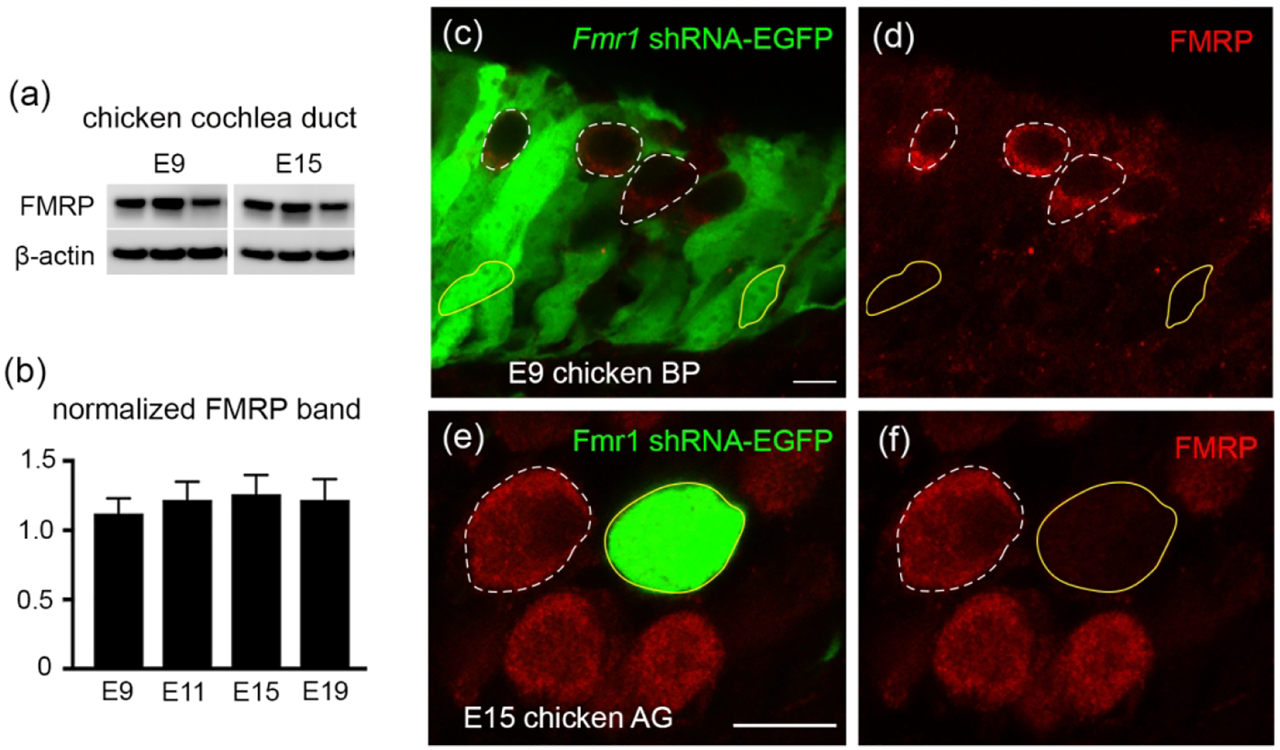
Antibody characterization for FMRP_PA8263 in the chicken inner ear. (a) Western blot of FMRP on cochlea samples collected from E9 and E15 chicken embryos. (b) Bar chart of FMRP band intensity that was normalized to β-actin and plotted with age (one-way ANOVA: F (3,9) = 0.6702; p = 0.5914). (c–d) FMRP immunostaining (red) on the E9 basilar papilla (BP) transfected with Fmr1 shRNA-EGFP (green). (e–f) FMRP immunostaining (red) on the E15 auditory ganglion (AG) transfected with Fmr1 shRNA-EGFP (green). Yellow solid and white dashed circles are examples of transfected (EGFP positive) and non-transfected (EGFP negative) cells or neurons, respectively. Scale bars = 10 μm in c, applies to c–d; 10 μm in e, applies to e–f.
