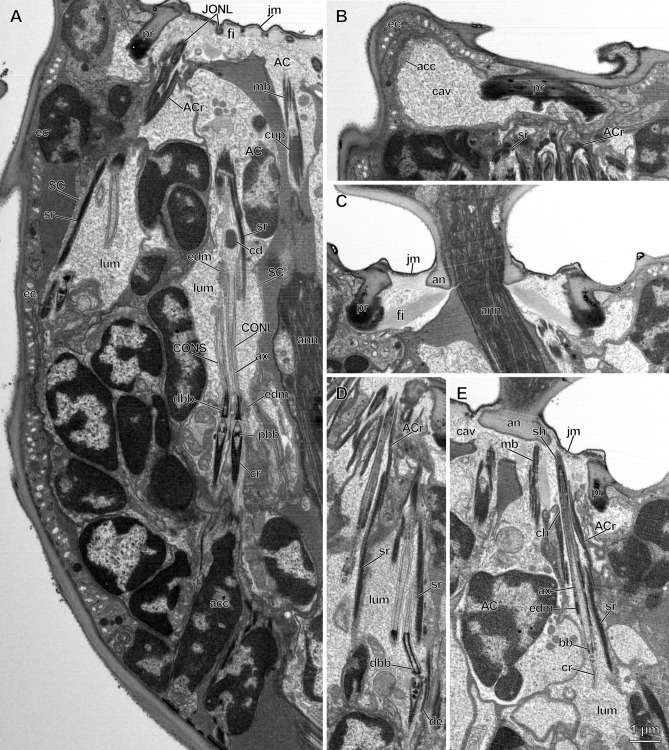
Figure 2.
Longitudinal sections of pedicellar chordotonal organs in Megaphragma viggianii. (A) Median section of pedicel, showing the dorsal scolopidia. Basal bodies, cilia roots and axonemes are visible in central organ (CO) cilia which are surrounded by two bands of electron-dense material. Cilium dilation of long CO neuron (CONL) encloses a core. Scolopale cell (SC) form lumina, scolopale rods and cups. Attachment cells (AC) of Johnston’s organ (JO) form AC rods, and AC of CO form microtubule bundles. Cilia tips of long JO neurons (JONL) run alongside the invaginations of joint membrane between fibers. (B) Parasagittal section of the distal part of the pedicel. Accessory cell forms cavity filled with granular material. (C) Median section of the distal part of the pedicel. A joint membrane and fibers connect the pedicel rim and annelus. (D) Longitudinal section through the distal part of ventral JO scolopidia. A distinct bend of short JO neuron (JONS) cilium is observed. (E) Parasagittal section through the distal part of the pedicel. JONL contains only one basal body, a small ciliary root and an axoneme distally tapering in a bundle of microtubules. Its sheath-covered tip attaches to the annelus alongside CO AC. A narrow channel formed by the JO AC connects the lumen and cavity. (acc—accessory cell, ACr—AC rods, an—annelus, ann—antennal nerve, ax—axoneme, bb—basal body, cav—cavity, cd—cilium dilation, ch—channel, cr—cilia root, dbb—distal basal body, de—desmosome, ec—epidermal cell, edm—electron-dense material, fi—fiber, jm—joint membrane, ju—junction, lum—lumen, mb—microtubule bundle, me—mesaxon, pbb—proximal basal body, pr—pedicel rim, sh—sheath, sr—scolopale rod).

