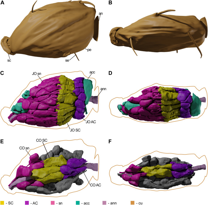
Figure 4.
3D models of M. viggianii pedicel, JO and CO. (A), (C), (E)—medial view, (B), (D), (F)—dorsal view. (A), (B): cuticular surfaces; (C), (D): JO scolopidia and accessory cells; (E) (F): CO scolopidia, half of the JO scolopidia models are absent; the present half is colored grey. (acc—accessory cell, an—annelus, ann—antennal nerve, cu—cuticle, pe—pedicel, sc—scape, se—sensilla, sn—sensory neurons).

