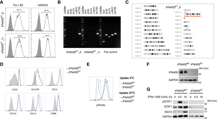
Figure 1.
A model of IFNAR2 deficient human iPS-macrophages (IFNAR2 PT iPS-Mϕ). (A) Expression of pluripotency markers by IFNAR2 PT iPSC clones 6 and 11 by flow cytometry.(B) PCR showing clearance of Sendai virus vector from IFNAR2 PT iPSC clones 6 and 11. (C) Karyogram produced from SNP array showing no gross abnormalities in the previously unpublished IFNAR2 PT iPSC clone 6. Red bars indicate loss or single copy, grey indicates loss of heterozygosity on chromosome 21 in the region of IFNAR2 (representative of data in IFNAR2 PT clone 11). (D) Expression of macrophage surface markers in IFNAR2 PT (clone 6) and IFNAR2 WT (WT1) iPS-Mϕ by flow cytometry, representative of repeat experiments in IFNAR2 PT clone 11 and WT2. (E) Phagocytic uptake of Zymosan pHrodo particles in IFNAR2 PT (clone 6) and IFNAR2 WT (WT1) iPS-Mϕ, representative of repeat experiments in clone 11 and WT2. (F) Immunoblot of IFNAR2 and GAPDH in IFNAR2 WT (WT2) and IFNAR2 PT (clone 11) iPS-Mϕ, representative of repeat experiments in WT1 and clone 6. (G) Immunoblot of IFN-I signalling in IFNα2b (1000 IU/mL) treated IFNAR2 PT (clone 11) and IFNAR2 WT (WT1) iPS-Mϕ, representative of n = 3 independent experiments.

