
Figure 2.
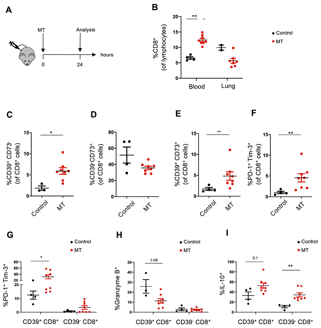
Circulating CD39+CD8+ T cells exhibit features of T cell exhaustion on treatment with liver-derived MT in vivo. (A) Wild-type recipient mice were injected intraperitoneally with liver-derived MT and studied 24 hours later. This MT dose reflects the expected amount of mitochondria released after damage of 20% of liver during major trauma. Blood was collected by cardiac puncture and processed for flow cytometry. (B) Frequencies of CD8+ T cells within peripheral blood-derived and lung-derived lymphocytes in control (untreated) and MT treated mice are shown. Frequencies of CD39+CD73− (C), CD39−CD73+ (D), double positive CD39+CD73+ (E) and PD-1+Tim-3+ cells (F) within blood-derived CD8+ T cells are represented along with the frequency of PD-1+Tim-3+ (G), granzyme B+ (H) and IL-10+ cells (I) among blood-derived CD39+CD8+ and CD39− CD8+ subpopulations. Data represent mean±SEM (n=4 in control group, n=8 in MT treated group). P value obtained using Mann-Whitney U test. *p<0.05; **p<0.01. IL, interleukin; MT, mitochondria; PD-1, programmed cell death 1.