Figure 3.

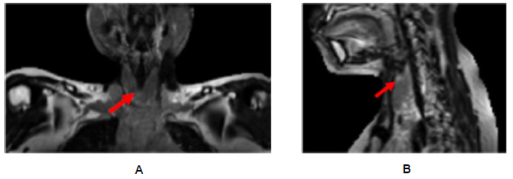
MRI of thyroid of the same patient, showing non-enhanced T2-weighted coronal (A) and sagittal (B) images with inhomogeneous high signal intensities within the thyroid parenchyma supportive of Hashimoto thyroiditis.

MRI of thyroid of the same patient, showing non-enhanced T2-weighted coronal (A) and sagittal (B) images with inhomogeneous high signal intensities within the thyroid parenchyma supportive of Hashimoto thyroiditis.