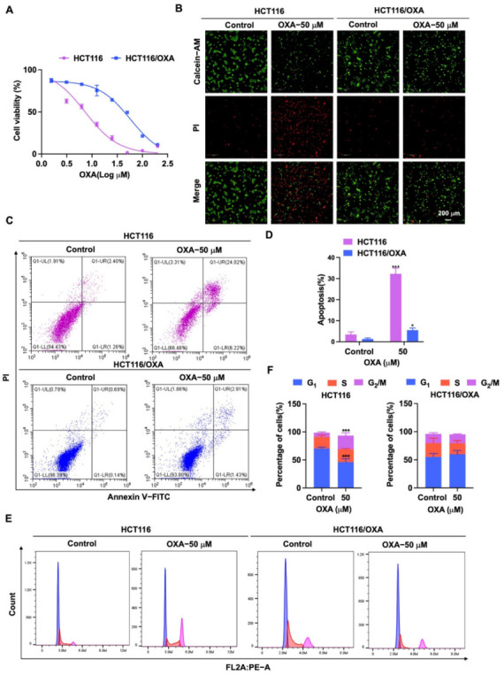
Figure 3.

Effect of OXA on the proliferation and growth of HCT116 and HCT116/OXA cells. (A) In CCK-8 experiment, HCT116 and HCT116/OXA were treated with different concentrations of OXA for 48 h; (B) OXA (50 μM) was applied to HCT116 and HCT116/OXA cells for 48 h, and Calcein-AM/PI staining was performed for live/dead cells; (C–F) HCT116 and HCT116/OXA cells were treated with OXA (50 μM) for 48 h to detect cell apoptosis and cell cycle progression (compared to the control group, * p < 0.05, *** p <0.001).
