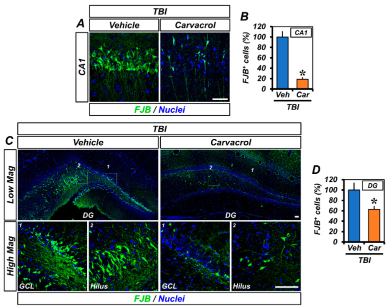
Figure 2.
Carvacrol reduced neuronal degeneration after TBI: (A,C) Representative images showing sections of the hippocampal CA1 (A) and GCL and hilus of DG (C) stained with FJB to detect degenerating neurons. Nuclei are stained with DAPI (blue). Scale bar, 50 µm; and (B,D) quantification of the number of FJB+ cells from the CA1 (B) and DG (D) of the ipsilateral hippocampus (mean ± SEM; n = 5–6 per group). * p < 0.05 vs. vehicle-treated TBI group (unpaired Student’s t-test).

