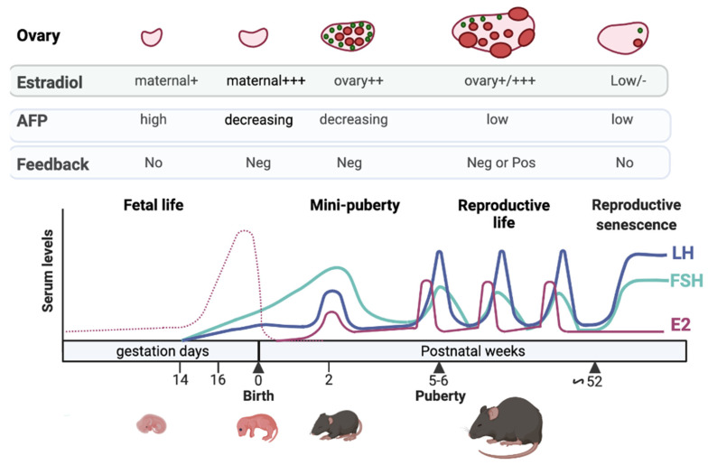
Figure 1.
Regulation of the gonadotrope axis at different periods of life in the mouse. FSH (green line) and LH (blue line) are detected in the last part of gestation, induced by GnRH stimulation. The high levels of maternal estrogens (dotted pink line) may not exert negative feedback at this time on FSH and LH synthesis and secretion due to high levels of α-fetoprotein (AFP) produced by the fetal liver. After birth, maternal estradiol wanes from pup serum, and this may lead to the loss of estradiol negative feedback and to the dramatic rise in gonadotropin levels observed at the time of mini-puberty. Additional ovarian factors may contribute to this rise (see the text). The gonadotropin surge contributes to increased estradiol production by the ovary at mini-puberty (continuous pink line). After that, the decrease in AFP after birth, and the maturation of inhibin negative feedback, may contribute to the observed fall in gonadotropin levels, remaining low up to puberty. During reproductive life, gonadotropin levels are regulated by both estradiol negative and positive feedback. Estradiol negative feedback in aged mice is attenuated, thereby increasing gonadotropin levels. Note that the levels of hormones shown during the different periods are given on an indicative basis since no studies encompass all these periods. See references in the text. The figure was prepared with the help of Mr. Le Ciclé, using BioRender, under Academic License terms.

