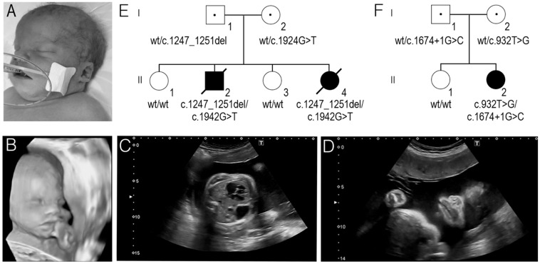
Figure 1.
(A) The facial view of the second affected newborn in family 1 (F1.II-4). (B–D). A 3D ultrasound image of the second foetus in family 1 (F1.II-4). Note: wide nasal bridge, broad nasal tip, wide mouth, short philtrum (B); Congenital diaphragmatic hernia (C); Wide mouth (D). (E) The segregation of the c.1942G>T and c.1247_1251del PIGN variants in family 1. (F) The segregation of the c.932T>G and c.1674+1G>C PIGN variants in family 2.

