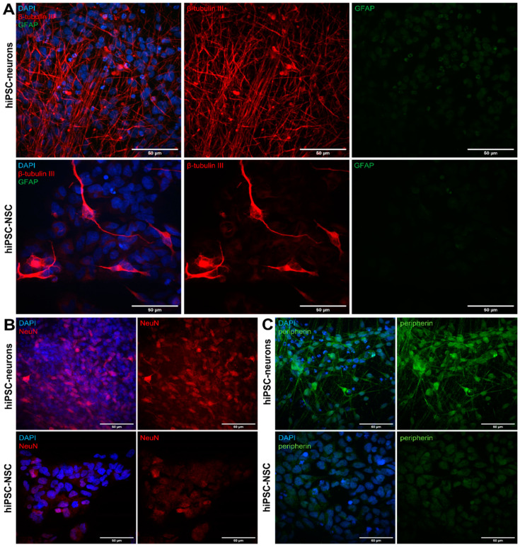
Figure 1.
Differentiation of hiPSC-NSCs into peripherin-expressing hiPSC neurons. (A) hiPSC-NSCs and hiPSC neurons were stained with mouse anti-β-tubulin III and rabbit anti-GFAP primary antibodies in combination with goat anti-mouse AF555 (red) and goat-anti rabbit FITC (green) secondary antibodies. Nuclei were stained with DAPI (blue). Representative images from three independent repeats of hiPSC-NSCs at DIV 2 and hiPSC neurons at 21 dpd. (B) hiPSC-NSCs and hiPSC neurons were stained with guinea pig anti-NeuN and secondary donkey anti-guinea pig Cy3 (red) antibody. Nuclei were stained with DAPI (blue). Representative images from three independent repeats of hiPSC-NSCs at DIV 2 and hiPSC neurons at 21 dpd. (C) hiPSC-NSCs and hiPSC neurons were stained with chicken anti-peripherin and secondary goat anti-rabbit FITC (green) antibody. Nuclei were stained with DAPI (blue). To verify specificity of the neuronal markers, secondary antibody controls were prepared for hiPSC-NSCs and hiPSC neurons (data not shown). Representative images from three independent repeats of hiPSC-NSCs at DIV 2 and hiPSC neurons at 21 dpd.

