Figure 2.
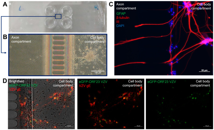
Generation of a compartmentalized neuronal culture model suitable for axonal VZV infection. (A) Image of a XonaChipTM (XC150). The two left wells and two right wells are separated from each other through a microgroove barrier (150 µm length, 10 µm width). (B) Brightfield image of a hiPSC-derived neuronal culture showing separation of cell bodies from axon termini. Representative image (at 18 dpd) with images that were acquired between 7 dpd and 21 dpd. (C) hiPSC-neuronal cultures were stained with mouse anti-β-tubulin III and rabbit anti-GFAP primary antibodies in combination with goat anti-mouse AF555 (red) and goat-anti rabbit FITC (green) secondary antibodies. Nuclei were stained with DAPI (blue). Representative image (at 13 dpd) with images that were acquired between 7 dpd and 21 dpd. (D) Spreading of eGFP-ORF23 VZV in the cell body compartment following inoculation of axon termini. Images show expression of eGFP, arising from the VZV capsid protein ORF23, and VZV gE which was stained with mouse anti-VZV gE primary antibody in combination with rabbit anti-mouse AF555 (red) secondary antibody. Representative image at 7 dpi (28 dpd).

