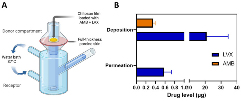
Figure 4.
Skin permeation and deposition results. (A) Schematic representation of the modified Franz cell setup for the ex vivo skin permeation and deposition study. The chitosan films containing AMB+LVX were placed on top of the full-thickness porcine skin and 10 µL of water was added between the film and skin to facilitate adhesion and mimic the moist environment of the wound tissue. The skin tissue was homogenized to extract the drug deposited and the samples from the receptor compartment were analysed for permeation data. (B) Ex vivo permeation and deposition results of AMB and LVX from the chitosan films. (Means + SD, n = 3).

