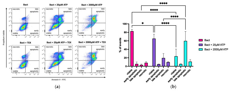
Figure 5.
Flow cytometry analysis of annexin V and propidium iodide (PI) staining in activated B cells exposed to ATP and/or TEX. (a) Adding ATP to activated B cells (Bact) induced apoptosis in B cells, especially the high 2000 µM ATP dose (upper panel). TEX (10 µg/mL) did not appear to alter apoptosis or necrosis (lower panel). FITC = fluorescein isothiocyanate. (b) Bar charts showing the influence of ATP and TEX on cell death in B cells by annexin V and PI staining. The mean of three independent experiments with standard deviation is presented. ATP significantly reduced the viability of activated B cells and pushed them towards apoptosis (viable: Bact vs. Bact + 20 µM ATP: p * = 0.0384; Bact vs. Bact + 2000 µM ATP: p **** < 0.0001; Bact + 20 µM ATP vs. Bact + 2000 µM ATP: p **** < 0.0001; late apoptotic: Bact vs. Bact + 2000 µM ATP: p **** < 0.0001; Bact + 20 µM ATP vs. Bact + 2000 µM ATP: p **** < 0.0001). Adding TEX to either ATP concentration did not significantly alter the results (Supplementary Figure S1). Statistical significance was determined using a two-way ANOVA with Tukey’s multiple comparisons test.

