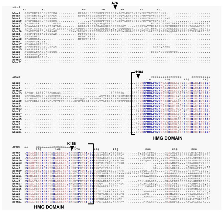
Figure 4.
Multiple sequence alignment of Sox family of proteins. The secondary structures are displayed on the top of the alignment for HMG domain. Identical residues are shown in blue, whereas similar residues are shown in red. We have omitted initial N- terminal and the C-terminal alignment portion from the final figure.

