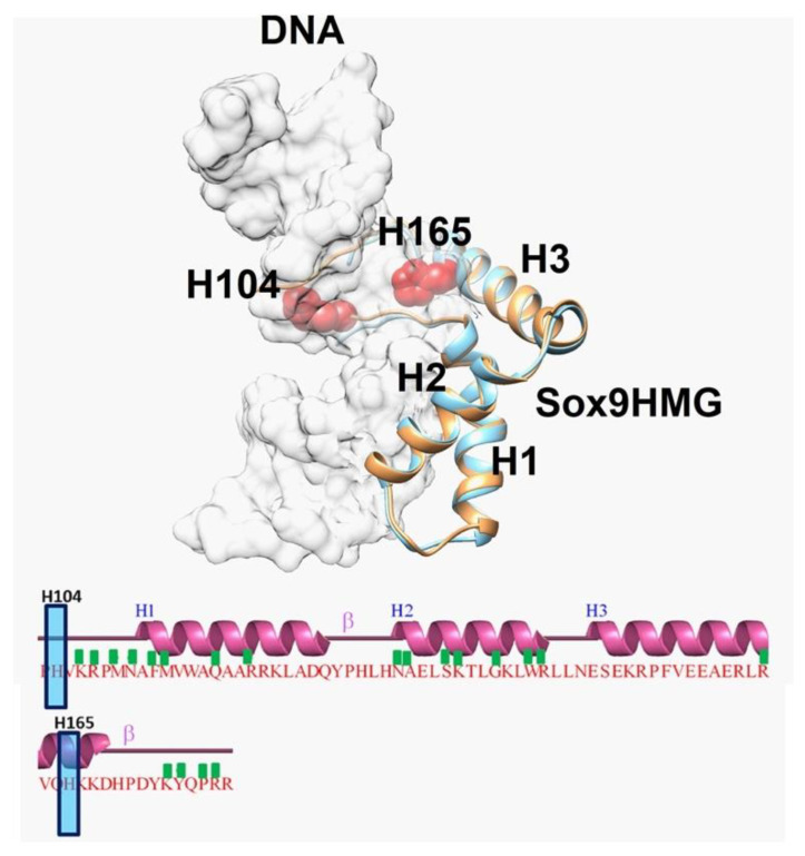
Figure 6.
Superposition of Sox9 structures showing key residues implicated in dimerization: Superposition of mouse Sox9HMG domain structure (PDBID: 4S2Q) in complex with DNA with human Sox9HMG domain structure in complex with DNA (PDBID: 4EUW). mSox9HMG is in brown, whereas hSox9HMG is in light blue. For clarity purposes, DNA is shown as light grey in transparent surface representation. The HMG domain contains three helices (H1, H2, and H3), and their interactions with DNA are represented with a green box. H104 and H165 are not part of the interaction and are shown as red.

