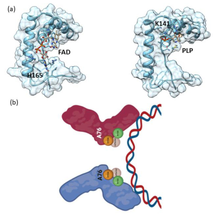
Figure 9.
In silico/docking Sox9 HMG-FAD/PLP interaction. (a) Cartoon as well as surface representation of the Sox9 HMG bound to FAD/PLP. Protein was rendered according to secondary structure elements. The complex structure highlighting the binding interfaces. (b) Schematic diagram of the cofactor binding to specific residues with the dimer formation leading to binding to DNA.

