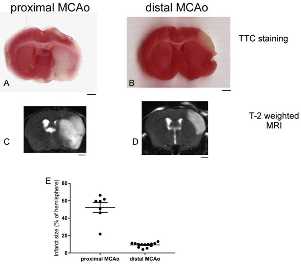
Fig. 1.

Proximal MCAo (pMCAo) and distal MCAo (dMCAo) induces different infarct size and locations in mouse brain. (A-B) TTC staining, (C-D) T2-weighted MRI images of pMCAo and dMCAo after 24 h. (E) quantification of infarct size using T2-weighted MRI imaging. The data are presented as MEAN± SEM (n = 7 for pMCAo and n = 13 for dMACo). Scale bar = 1 mm.
