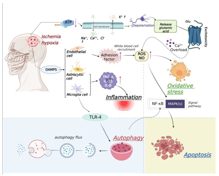
Figure 2.
Relationship between ischemic stroke and apoptosis, inflammation, autophagy, and oxidative stress. After cerebral ischemia, the activity of Na+/K+−ATP enzyme in ischemic penumbra decreases and the imbalance of ion homeostasis leads to cell membrane depolarization and Ca2+ influx, resulting in excessive release of glutamate and excitotoxicity. With the influx of a large amount of Ca2+, the adhesion factors of endothelial cells increase, which leads to white blood cell recruitment, increases the contents of ROS and nitric oxide (NO), and causes oxidative stress. Then, NF-κB and MAPK signaling pathways are activated, causing cell apoptosis. Microglial and astrocytic cells are also activated, which up-regulate tumor necrosis factor (TNF-α), interleukin-1β (IL-1β), and interleukin-6 (IL-6), finally causing an inflammatory reaction. Meanwhile, microglia cell-mediated down-regulation of Toll-like receptor-4 (TLR-4) induce autophagy.

