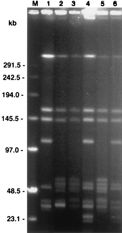
FIG. 3.
PFGE of MluI digests of B. burgdorferi isolates and subclones of isolate 225c. Lanes: 1, isolate WFMC4 (PFT A); 2, B31 (PFT B); 3 and 4, P. leucopus isolates 225a and 225c, respectively; 5 and 6, isolate 225c subclones 2E7′ and 3B6′, respectively; M, DNA pulse marker, 0.1 to 200 kb (Sigma). The gel photograph was scanned into Adobe Photoshop 3.1, scaled to the column width, and labeled.

