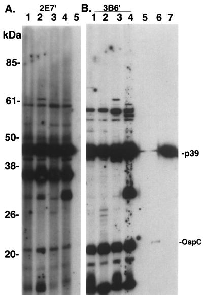
FIG. 6.
Immunoblot analysis of sera from mice in the superinfection experiment. Serum (diluted 1:150) from mice infected with either subclone 2E7′ (lanes 1 to 4 in panel A) or 3B6′ (lanes 1 to 4 in panel B) were reacted against the homologous B. burgdorferi subclone antigens (2E7′ in panel A and 3B6′ in panel B). Serum from an uninoculated mouse is included in lane 5 of each panel. Lanes 6 and 7, MAbs to B. burgdorferi-specific antigens ospC and p39, respectively. All sera were detected with POD-labeled secondary antibody (BM Chemiluminescence; Roche). Seroreactivity at approximately 30 kDa in panel A probably corresponds to OspD expression in 2E7′, which is encoded on the 38-kb plasmid (subclone 3B6′ lacks plasmids at 36 and 38 kb). Prestained molecular mass markers (Life Technologies) are indicated on the left. The immunoblots were scanned into Adobe Photoshop 3.1, scaled to the column width, and labeled.

