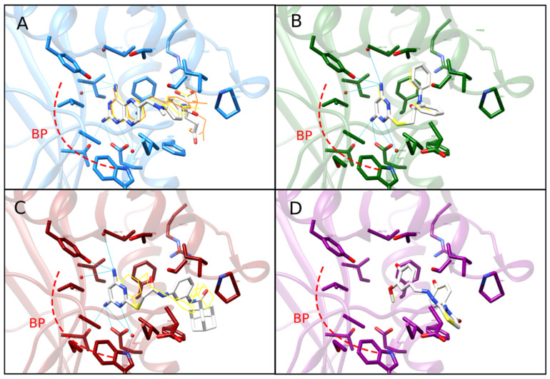
Figure 3.
Predicted binding mode of compounds C1 and C2 on human DHFR active site. Superimposed 3D docked poses of the inhibitor MTX (A), hit compounds C1 (B) and C2 (C), and an hDHFR decoy (D) interacting with the hDHFR active site. In each case, ligands from initial docking are displayed in a yellow wire representation, and those from re-docking (i.e., including a structurally conserved water molecule) are shown as white sticks colored by heteroatom. The structure of hDHFR is represented as ribbons and the side chains of critical active site residues are shown as sticks colored by heteroatom. The original pose for MTX (PDB code: 1U72) is represented as an orange wire (see panel (A)). H-bonds are shown only for re-docking poses and are displayed as blue continuous lines. The main binding pocket (BP site) of DHFR is illustrated in all panels as a red discontinuous curved line.

