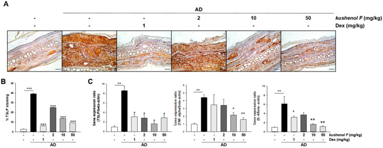
Figure 4.
TSLP and cytokine expression analysis of dorsal skin lesions in DFE/DNCB-treated atopic dermatitis mice treated with kushenol F. (A) Paraffinized ear sections were examined using the TSLP antibody. Scale bar = 10 µm. (B) TSLP expression levels were quantified using ImageJ software. (C) TSLP, TNF-α and IL-4 mRNA expression levels in the ear skin of kushenol F-treated mice. Data are presented as the mean ± SD (n = 5). * p < 0.05, ** p < 0.01 and *** p < 0.001 vs. control. TSLP, thymic stromal lymphopoietin; DFE, Dermatophagoides farinae extract; DNCB, 1-chloro-2,4-dinitrochlorobenzene.

