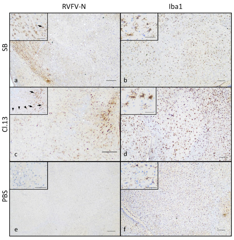
Figure 4.
Brain of SB- and Cl.13-IN-infected mice displays N-protein-positive cells and activated microglia. Mice intranasally infected with the SB (a,b) or Cl.13 (c,d) RVFV strains, or inoculated with PBS (e,f) were euthanized at day 6–10 (Cl.13) or day 5–7 (SB) p.i. when moribund, or day 20 post-inoculation (PBS) and their brain was analyzed by immunohistochemical staining against RVFV N (a,c,e) and Iba1 (b,d,f) proteins. Representative low magnification (×4) images show immunoreactivity to RVFV N-protein in the cranioventral portion of the brain of SB-infected mice (a) and the midbrain of Cl.13-infected mice (c) and compared to uninfected controls (e). Higher magnification insets (×20) show cells with morphological features of neurons (arrows) and glial cells (arrowheads). Microglial reactivity assessed by Iba1 staining was mild in SB-infected mice (b) and more pronounced in Cl.13-infected mice with activated microglial cells ((d), inset). Bars: 200 µ in low magnification images and 50 µ in high magnification insets.

