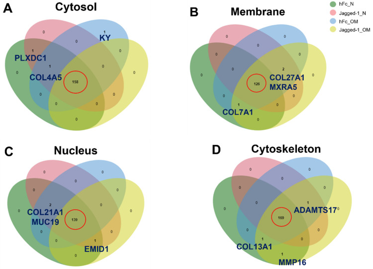
Figure 5.
Venn diagram of common proteins and unique proteins presented in the (A) cytosol, (B) membrane, (C) nucleus, and (D) cytoskeleton compartment, respectively. The analyses were performed using an interactive Venn diagram viewer [31]. The red circle indicated the common matrisome proteins of the different culture conditions.

