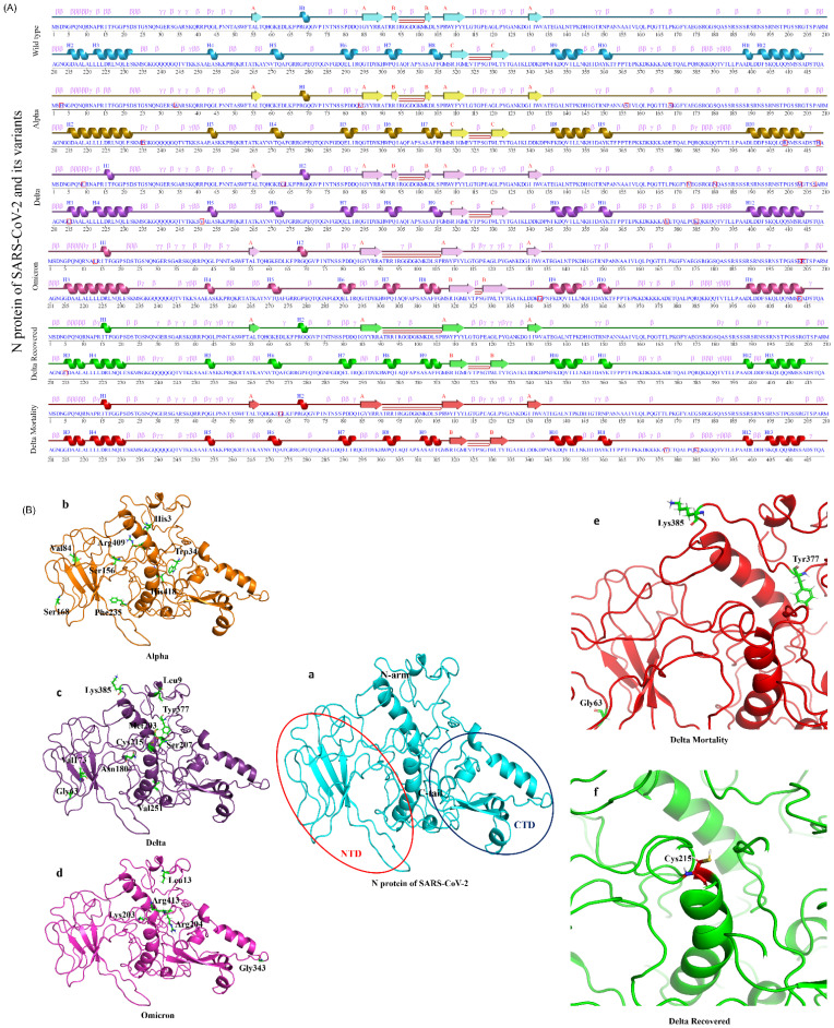
Figure 6.
2D and 3D structural changes in the N protein with respect to the variants and their clinical outcomes. (A) Secondary structure representation of the targeted N protein and its variants, along with the “□” represents the mutated amino acid(s). (B) Molecular modeling of the N protein wild type (a) (Red circle represents the NTD domain and the dark blue color represents the CTD domain), Alpha (b), Delta (c), Omicron (d), DM (e), and DR (f).

