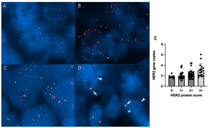
Figure 4.
(A) HER2 diploid in canine mammary carcinoma: two gene copies of both HER2 (red signal) and CRYBA1 (green signal), FISH. (B) HER2 amplification in canine mammary carcinoma: HER2/CRYBA1 > 2, FISH. (C) HER2/CRYBA1 co-amplification/polysomy: increased copy numbers of both HER2 and CRYBA1 (> 4 gene copies). (D) HER2/CRYBA1 fusion pattern: close co-localization of HER2 and CRYBA1, interpreted as translocation and fusion of the two genes (arrows) together with non-fused alleles in the same nuclei (arrowheads). (E) Histogram representation of the correlation between HER2 overexpression with an increased number of HER2 gene copies (p = 0.01, R = 0.24; Spearman test).

