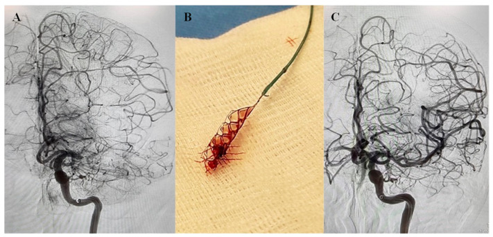
Figure 1.
Cerebral Angiogram of acute stroke with left middle cerebral artery (MCA) occlusion. (A) Cerebral angiograph showing left MCA M1 occlusion. (B) After two passes with Solitaire (4 × 20 mm), vessel recanalizes (TICI 3). (C) Post-thrombectomy angiogram showing complete recanalization of the left MCA.

