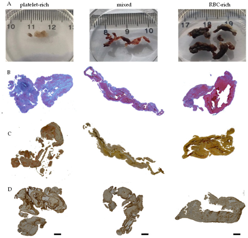
Figure 2.
Distinct red blood cell (RBC)-rich and platelet-rich areas are seen in stroke thrombi. (A) Representative photomacrographs of the platelet-rich, mixed, and RBC-rich thrombi were retrieved from ischemic stroke patients. (B) Martius Scarlet Blue (MSB) staining of the three representative thrombi sections. Fibrin-rich areas appear red while RBC-rich areas appear yellow. (C) Hematoxylin and Eosin (H and E) staining depicting RBC-rich areas (red) and RBC-poor areas (light pink). (D) Immunohistochemical staining shows von Willebrand factor (VWF) in the three representative thrombi sections. Scale bars (B–D) = 500 μm.

