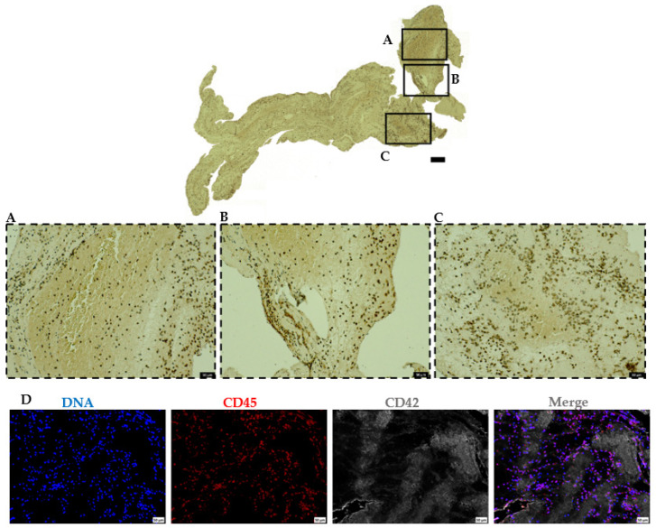
Figure 6.
Abundant leukocyte accumulation mainly at the interface between platelet-rich and RBC-rich areas. (A–C) Immunohistochemical staining of retrieved AIS patient thrombi to visualize leukocytes using CD45 antibody (upper panel). Higher magnification of the representative staining is shown in inserts (lower panels). Scale bars: 200 μm (top panel) and 50 μm (lower panels). (D) Representative ischemic stroke patient thrombi stained for leukocytes (CD45, red), platelets (CD42b, grey), and DNA (DAPI, blue). Scale bars: 50 μm.

