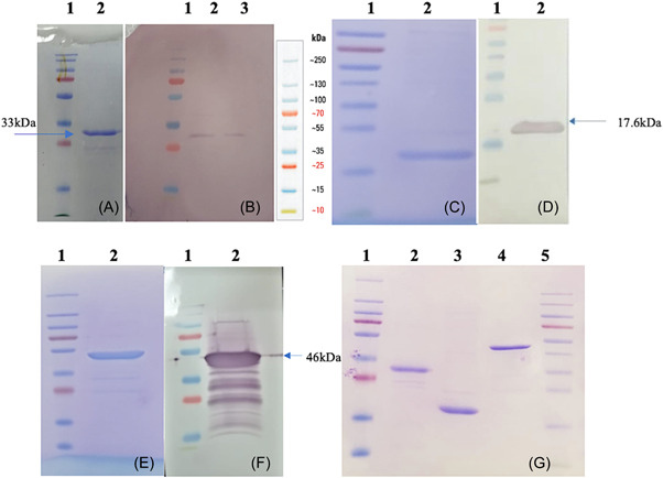
Figure 1.

(A) SDS‐PAGE detection of the SARS‐CoV‐2 spike protein S1 region fragment protein (P1:33 kDa) containing RBD; Lane 1, 2: protein ladder and purified P1 protein elute. (B) WB analysis of P1 protein. WB was done using an anti‐Spike‐RBD‐mIgG2a‐monoclonal mouse IgG2a antibody (Invivogen). (C) SDS‐PAGE detection of the SARS‐CoV‐2 spike protein S2 region fragment protein (P2: 17.6 kDa); Lane 1, 2: protein ladder and purified P2 protein elute. (D) Western blot analysis of P2 protein. WB was done using the serum obtained from mice immunized with combined recombinant P1 + P2 + N formulation adjuvanted with alum. (E) SDS‐PAGE detection of the SARS‐CoV‐2 nucleocapsid protein (N; 46 kDa); Lane 1, 2: protein ladder and purified N protein elute. (F) Western blot analysis of N protein. WB was done using an anti‐Nucleocapsid‐mIgG1‐monoclonal mouse IgG1 antibody (Invitrogen). (G) Profiles of purified P1, P2, and N proteins on SDS‐PAGE. RBD, receptor‐binding domain; SARS‐CoV‐2, severe acute respiratory syndrome coronavirus 2; WB, Western blot.
