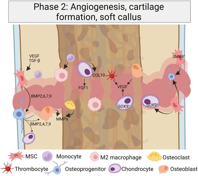
Figure 2.
Intramembranous ossification and endochondral ossification occur in parallel, but with different underlying mechanisms. Whereas periosteal mesenchymal stem cells (MSCs) adjacent to the fracture site form a hard callus via intramembranous ossification, the central area with a hypoxic core forms a soft callus via endochondral ossification. The macrophages are M2-polarized and alternatively activated (AAM), and the role of chondrocytes in the soft callus development becomes more prominent. Created with BioRender.com.

