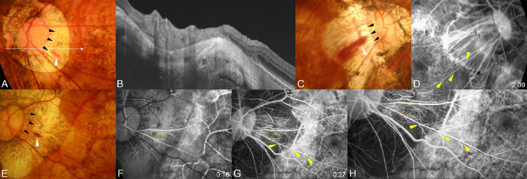
Figure 3.
Hemorrhages present at the peak of a scleral ridge and a straightened segment of retinal arterioles running away from the optic disc and telangiectasia of the surrounding capillaries. (A) Fundus photograph of the left eye of a 55-year-old woman with an axial length of 31.42 mm showing a typical flame-shaped conus hemorrhage (white arrowhead). The peak of the ridge can be seen (black arrowheads). The long arrow indicates the OCT scan line for the image shown in B. (B) OCT image showing a prominent scleral ridge at the hemorrhagic site. (C) Fundus photograph of the right eye of a 68-year-old woman with an axial length of 31.19 mm showing a typical flame-shaped conus hemorrhage (white arrowhead). The peak of the ridge can be seen at one end of the hemorrhage (black arrowheads). (D) Fluorescein angiogram taken one year before the hemorrhage at two minutes after the dye injection showing a marked bending of the retinal venules on the straightened artery (arrowheads). (E) Fundus photograph of the left eye of a 71-year-old woman with an axial length of 31.78 mm showing a typical flame-shaped conus hemorrhage (white arrowhead). The peak of the ridge can be seen (black arrowheads). (F, G) Fluorescein angiograms taken one year before the hemorrhage at 16 seconds after the dye injection (F) and at 27 seconds after dye injection (G). The hemorrhage site is circled in yellow. A marked bending of the retinal venules can be seen especially at the A/V crossing sites of the straightened artery (arrowheads). The closer the bending site is to the optic disc, the sharper is the bending angle. Many retinal microaneurysms and retinal capillary telangiectasias can be seen in the temporal part of the conus. (H) Magnified image shows capillary telangiectasia and microaneurysms.

