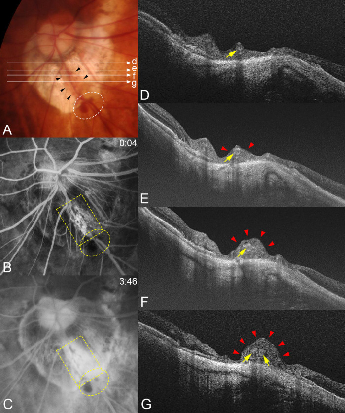
Figure 4.
Compression of the retinal tissue and dye leakage from the compressed area in a patient with a periconus hemorrhage. (A) Fundus photograph of the left eye of a 74-year-old woman with an axial length of 29.80 mm showing periconus hemorrhage (white circle) with a blotchy shape. The fundus color is slightly orange in the area along the lower temporal retinal vein (arrowheads) crossing the conus area. Long arrows indicate OCT scan lines for images in D, E, F, and G. (B) Early phase of FFA at four seconds after the dye injection shows an area with many capillaries with microaneurysms along both sides of this lower temporal retinal vein (yellow square). Hemorrhages are present along the vessels (yellow circle). (C) At three minutes after the dye injection, a massive dye leakage is seen from the dilated capillaries in B. Periconus hemorrhages show blocked fluorescence (yellow circle). (D–G) OCT images corresponding to the scan lines shown in A. Images show that the retinal tissue is severely compressed (red arrowheads) especially at sites farther away from the optic disc along the vessel (arrows). The retina around the compressed area is thin, especially in G.

