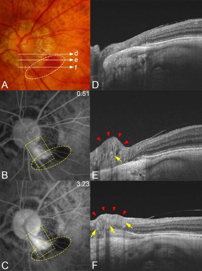
Figure 5.
Typical case showing a compression of the retinal tissue in a patient with a periconus hemorrhage. (A) Fundus photograph of the left eye of a 76-year-old woman with an axial length of 28.97 mm showing a blotchy shaped periconus hemorrhage (white circle). Long arrows indicate the OCT scan lines for images in D, E, and F. (B, C) Fluorescein angiograms. At 51 seconds after the dye injection showing leakage along the lower temporal retinal artery (yellow square) and hemorrhage developed adjacent to the margin of the myopic conus. Corresponding blocked fluorescence can be seen (yellow circle). (D, E) OCT images obtained two years after the hemorrhage of scan lines in A showing that the retinal tissue is severely compressed (red arrowheads) at the middle (E) in the area of conus from the optic disc along the retinal artery (arrow). (F) Three vessels (arrows) can be seen in the compressed retina (arrowheads).

