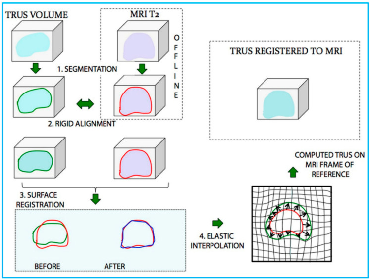
Figure 9.
Schematic illustration of the process of US/MRI fusion. MR and transrectal US (TRUS) images were (1) segmented and then (2) rigidly aligned. Fusion then proceeded, involving (3) a surface registration, and (4) elastic (non-rigid) interpolation. Reprinted from [135] with permission.

