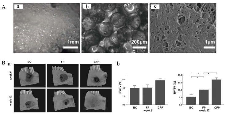
Figure 8.
The FP scaffold for bone repair. (A) Morphology and structure of FP scaffold. (a) Stereomicroscopy image of FP scaffold. (b) Image of FP scaffold under an inverted microscope. (c) SEM image of FP scaffold. (B) Micro-CT evaluation of in vivo bone growth within harvested mandible samples. (a) Three-dimensional reconstructed images at week 6 and 12. (BC: Blank control, bone defect without implant; FP: Bone defect implanted with FP scaffold; CFP: Bone defect implanted with cell-laden FP scaffolds). (b) Results of quantitative analyses of samples harvested at weeks 6 and 12. The symbol * denotes a statistical significance between two groups (p < 0.05). Adapted with permission from Ref. [82]. Copyright 2022 NPG Asia Materials.

