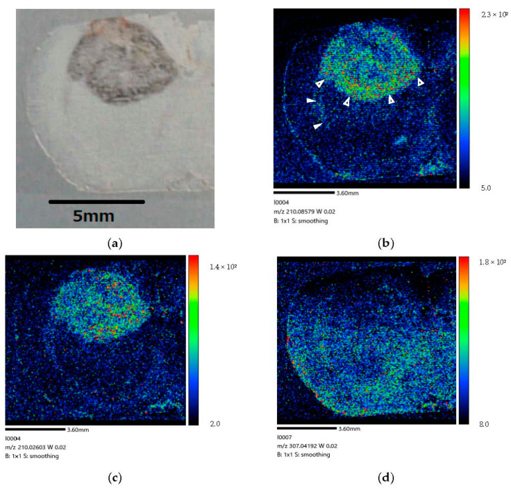
Figure 6.
(a) Coronal section of brain-tumor-model rat after 160 min administration. (b) BPA biodistribution image generated by MALDI-MSI. BPA penetration is mainly visible in the brain tumor tissue. Some BPA is present in the lateral ventricle (➣). BPA is localized in the invasive margin (➤) of the brain tumor and clearly distinguishes the border between tumor and normal cells. These results imply that BPA penetrated brain tumor cells within 160 min following administration into the lateral ventricle of brain-tumor-model rat. (c) The mass image obtained from peaks at m/z 210.026 originated in tumor. The image showed a distribution different from the localization of BPA within the tumor. (d) The mass image obtained from peaks at m/z 307.042 originated from endogenous biomolecule.

