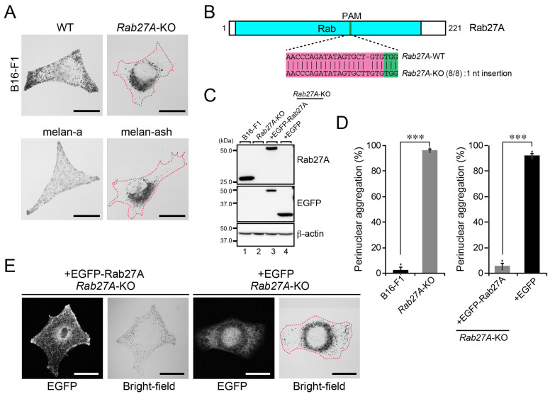
Figure 3.
Rab27A is essential for the peripheral distribution of melanosomes. (A) Representative bright-field images of WT B16-F1 cells, Rab27A-KO B16-F1 cells, melan-a cells, and Rab27A-deficient melan-ash cells. Cells showing perinuclear melanosome aggregation are outlined with broken red lines. Scale bars, 20 μm. (B) Domain organization of the mouse Rab27A protein and genomic mutations in Rab27A-KO cells. The Cas9 target and PAM sequences are highlighted in magenta and green, respectively. Genomic PCR products containing the target site were subcloned into the pGEM-T Easy vector. Then, eight colonies (denominators of the fractions in parentheses) were picked up at random and their inserts were sequenced. (C) Expression of Rab27A in WT cells and Rab27A-KO B16-F1 cells. Lysates of the cells indicated were analyzed by immunoblotting with antibodies against Rab27A (top), GFP (middle), and β-actin (bottom). (D) Quantification of the numbers of WT cells, Rab27A-KO cells, Rab27A-KO cells expressing EGFP-Rab27A, and Rab27A-KO cells expressing EGFP with perinuclear melanosome aggregation shown in (A,E). The graphs show the means and SEM of the data obtained in three independent experiments (n > 24 cells in each experiment). ***, p < 0.001 (Student’s unpaired t-test). (E) Representative bright-field images of Rab27A-KO cells expressing EGFP-Rab27A or EGFP alone. The EGFP-expressing cell is outlined with a broken red line. Scale bars, 20 μm.

