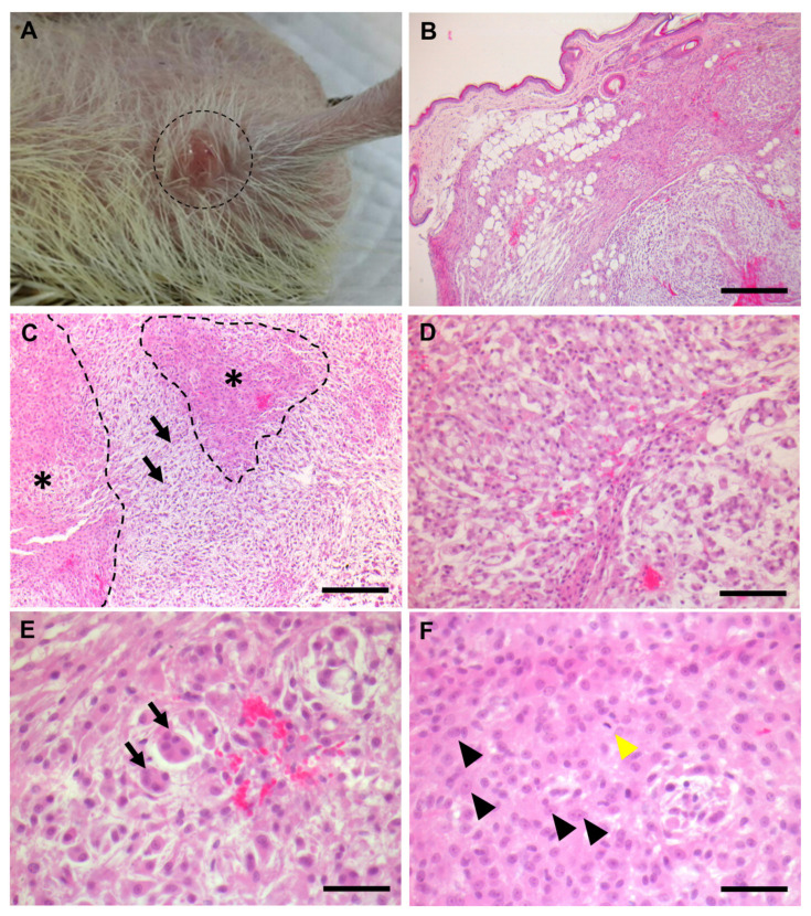
Figure 1.
Pathological observation of the pleomorphic myxoid liposarcoma. (A) Gross observation with gross pathology. The neoformations were protrude and dome-shaped. (B) Microscopic observations of low magnification. The neoformations were located in the subcutaneous layer of the right abdomen. The neoformations were not encapsulated. Scale bar = 500 μm. (C–F) Microscopic observations of high magnification. (C) The neoformations were composed of the myxoid proliferation area (black arrows) and cellular area (outlined and marked with asterisks). The myxoid proliferation area is composed of proliferating stellate cells and abundant myxoid materials. Scale bar = 200 μm. (D) Neoplastic adipoblasts with variable size of intracytoplasmic clear vacuoles. Scale bar = 100 μm. (E) Large bizarre multinucleated giant cells (arrows). Scale bar = 50 μm. (F) Mitotic figures were sometimes observed (yellow arrowhead). Epithelioid cells were observed (black arrowheads). The epithelioid cells had abundant eosinophilic cytoplasm with round-to-oval-shaped nuclei. One to two prominent nucleoli were observed in the nucleus. Scale bar = 50 μm.

