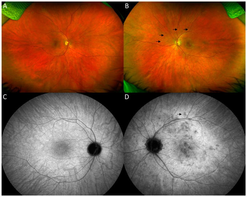
Figure 1.
Optos ultrawide-field fundus images of the right eye showing normal fundus (A). The left eye demonstrates a mottled appearance of the macula and areas of multiple scattered hypopigmented spots in the posterior pole (dark arrows) (B). Late-phase indocyanine green angiography (ICG) of the left eye shows multiple spots of hypocyanescence in the posterior pole, some of which correspond to the spots on color fundus photos (D). Late-phase ICG of the right eye (C) was unremarkable.

