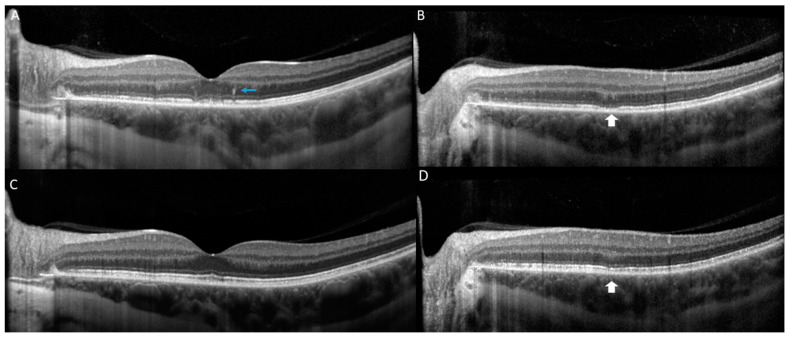
Figure 2.
Optical coherence tomography (OCT) of the left eye at the initial presentation is notable for presence of hyperreflective dots in outer retina (blue arrow) and multifocal loss of the ellipsoid zone (EZ) (white arrow) (A,B). At 1 month follow up, reconstitution of the EZ (white arrow) is observed and improvement in outer retinal hyperreflectivity (C,D).

