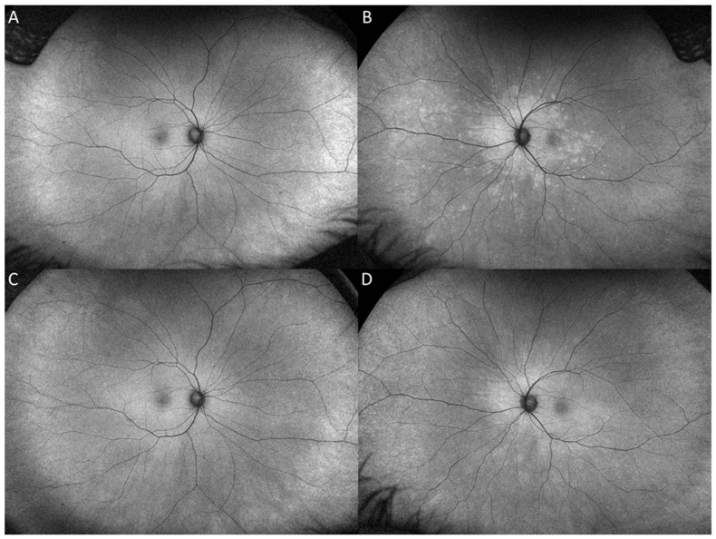
Figure 3.
Fundus autofluorescence (FAF) at presentation of the left eye at presentation demonstrates a confluent area of hyperautofluorescence surrounding the optic nerve and posterior pole with hyperautofluorescent dots extending to the temporal macula and mid nasal periphery (B) FAF at 1 month follow-up demonstrates decreased hyperautofluoresence and ill-defined borders. (D). FAF of the right eye at presentation and final visit are unremarkable (A,C).

