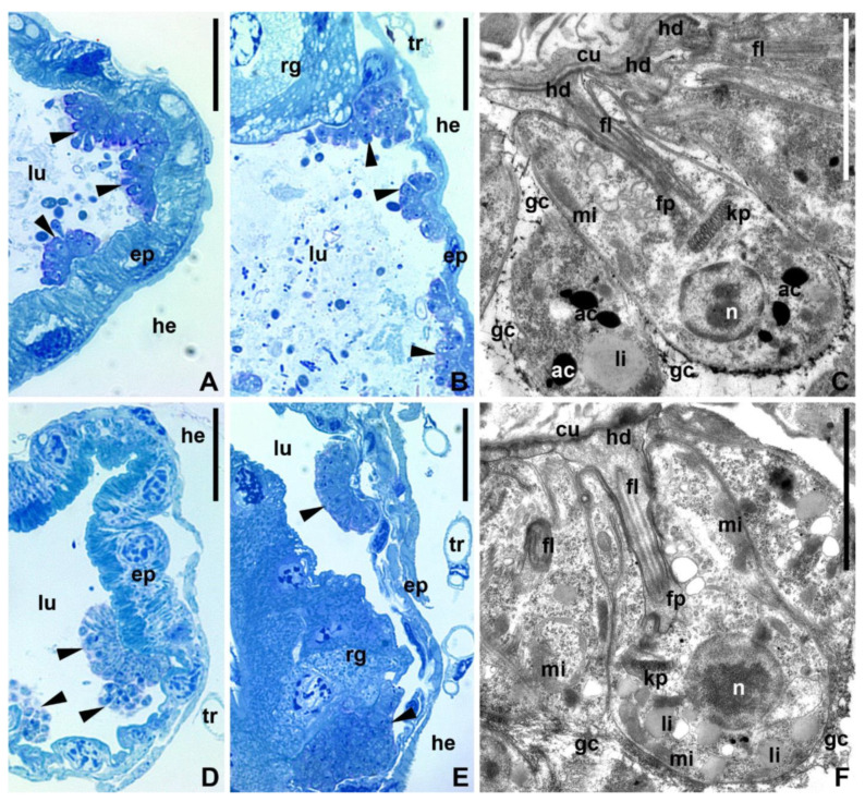
Figure 4.
Trypanosomatids in the hindgut of experimentally infected mosquitoes (96 h). (A,B,D,E)—semithin sections stained with methylene blue-azure stain (light microscopy); (C–F)—TEM. (A–C)—C. brevicula; (D–F)—C. dobrovolskii; (A,C,D,F)—ileum, (B–E)—rectum. Ac—acidocalcisome; cu—cuticle; ep—epithelium fl—flagellum; fp—flagellar pocket; gc—glycocalyx; hd—hemidesmosome; he—hemocoel; kp—kinetoplast; li—lipid droplets; lu—intestinal lumen; mi—mitochondrion; n—nucleus; rg—rectal gland; tr—trachea; arrowheads—attached parasites. Scale bars: (A,B,D,E)—20 µm; (C–F)—2 µm.

