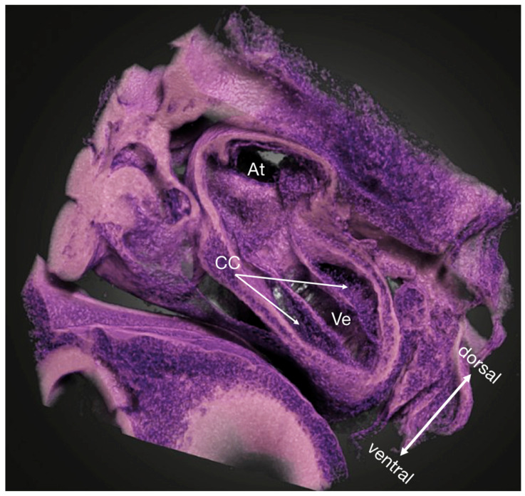
Figure 1.
Micro-CT volume rendering of a PbOAc stained HH18 chicken embryo showing a near-histological 3D view into the ventricle and the atrium (arbitrary cutaway). The cardiac cushions are in the process of being populated by cells. Owing to the high specificity of the PbOAc staining, the lookup table and the transfer function could be manipulated as to resemble the classic Hematoxylin/Eosin-staining used in histology to locate nuclei more easily. At atria, CC cardiac cushions, Ve ventricle.

