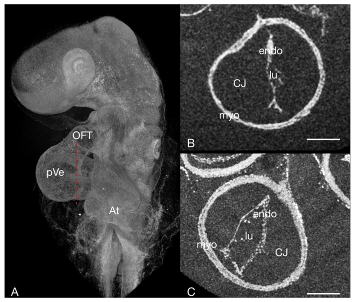
Figure 4.
The embryonic heart tube of a HH13 chick stained with PbOAc. (A) Overview, micro-CT volume rendering: the red dotted line corresponds to the sectioning plane of (B,C) Virtual cross-section through the embryonic heart tube in the area of the primitive ventricle fixed at different phases in the heart cycle. (B) systole—the lumen is slit-shaped; (C) diastole—the lumen appears more oval in shape. At atrium, CJ cardiac jelly, endo endocardium, lu lumen, myo myocardium OFT outflow tract, pVe primitive ventricle; scale bar = 100 µm.

