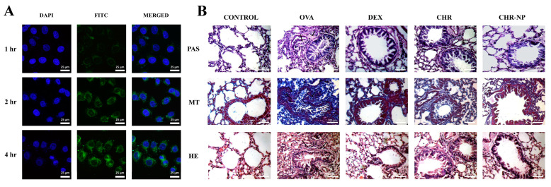
Figure 5.
(A) Immunofluorescence images of A549 cells. (B) Exemplar photomicrograph of lung tissues from all the experimental groups stained by periodic acid-Schiff (PAS), Masson’s trichrome (MT), and hematoxylin-eosin (H&E) staining. Reproduced with permission from [49], © 2022 Elsevier Inc.

