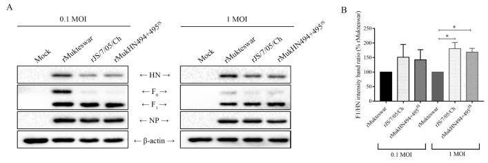
Figure 6.
Evaluation of F protein cleavage activities of NDVs bearing different HN proteins. (A) Three viruses were inoculated into CEF cells at 0.1 and 1 MOI, respectively. The amounts of HN, F0, and F1 proteins were detected by Western blotting and then the cleavage-promotion activity of different HN proteins was analyzed. (B) The gray bands were evaluated by the ImageJ software. The F1/HN ratio at each infected group was normalized to that of the rMukteswar group. * p < 0.05.

