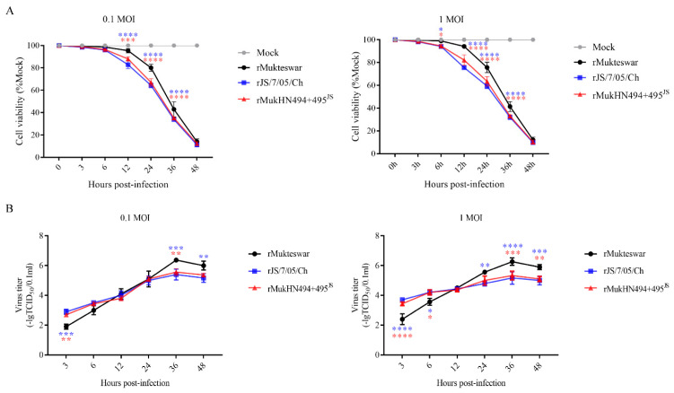
Figure 7.
Evaluation of cell viability and virus replication ability in NDV-infected CEF cells. Three viruses were inoculated into CEF cells at 0.1 and 1 MOI, respectively. (A) Reduction of cell viability in NDV-infected CEF cells at 3, 6, 12, 24, 36, and 48 hpi. (B) Growth characteristics of the three viruses were determined during 3–48 hpi. All values were compared with that of rMukteswar. * p < 0.05, ** p < 0.01, *** p < 0.001, and **** p < 0.0001.

